Osteoporosis

FREE READ
+How to treat
In print
FREE READ

Osteoporosis

By David Kim
32 minutes to Read
Osteoporosis
[Image: Fraser Williamson]

Primary care has a central role in identifying, evaluating and managing patients with osteoporosis and high risk of fracture. This article, written by David Kim, reviews the condition, patient risk assessment and the use of established and new treatments

This How to Treat has been endorsed by the RNZCGP and has been approved for up to 1 credit for continuing professional development purposes (1 credit per learning hour). To claim your CPD credits, complete the assessment at the end of this page, then log in to your Te Whanake dashboard and record this activity under the appropriate learning category.

Nurses may also find that reading this article and reflecting on their learning can count as a professional development activity with the Nursing Council of New Zealand (up to 1 PD hour).

Learning objectives

At the end of this course, you should be able to:

  • Discuss the role of primary care in managing osteopaenia, osteoporosis and fragility fractures

  • Explain how risk for osteoporosis and fragility fracture is determined

  • Describe the indications, contraindications and adverse effects of anti-osteoporosis medications

  • Discuss the follow-up of patients on anti-osteoporosis treatment

Domains of general practice

This How to Treat may assist with the development of the following domains of competence in the general practice curriculum:

  • Domain 1: Communication
  • Domain 2: Clinical expertise
  • Domain 3: Professionalism
  • Domain 4: Scholarship
  • Domain 5: Context of general practice
  • Domain 6: Management
Would you prefer to download this article?

This publication has been reprinted with the support of Osteoporosis New Zealand to provide an update on the management of osteoporosis in primary care. The content is entirely independent and based on published studies and the author’s opinion.

Download PDF

Osteoporosis is a chronic condition characterised by brittle bones with reduced bone mineral density leading to increased risk of fragility fractures, defined as fractures sustained from low-impact trauma (eg, a fall from standing height or less).

Bone mass reaches its peak in young adulthood and gradually declines beyond middle age. It is, therefore, not surprising that incidences of osteoporosis and fragility fractures are rising with our ageing population.

It is estimated that well over 20,000 New Zealanders experience one or more fragility fractures each year. With these fractures, there is tremendous individual suffering, such as pain, temporary or permanent loss of independence, and even increased mortality.

Fragility fractures also incur a huge and ever-increasing burden on our already strained healthcare system. ACC data from 2020 estimated that falls and fracture-related injuries among New Zealanders aged 65 and over cost $195 million per year, representing a 47 per cent increase since 2013. ACC estimates that “doing nothing” will more than double the cost burden by 2035 (tinyurl.com/ACC-prevent-FF).

With appropriate screening for, and management of, osteoporosis, a significant proportion of fragility fractures can be prevented.

The vast majority of those who experience fragility fractures are, at least in part, assessed and managed in primary care. Even if the initial care around the incident fracture is delivered by local secondary services, further follow-up and management of related issues (eg, pain, return to normal activities and future fracture prevention interventions) are often dealt with in primary care.

With widespread implementation of Fracture Liaison Services (FLS) in all regions of New Zealand, a large proportion of fragility fracture cases are now being identified and assessed in a timely fashion, with appropriate management strategies implemented or recommended. This secondary fracture prevention programme, mostly based in secondary settings, cannot function without cooperation and coordination with primary care for its care delivery, including patient investigation, initiation and continuation of pharmacotherapy and implementation of falls prevention.

In those not known to have osteoporosis or never having had a fragility fracture, screening for osteoporosis and treating the appropriate population can lead to early diagnosis and fracture prevention.

Osteoporosis and fragility fractures occur in a reasonably predictable manner in terms of demographics and clinical background. Those deemed to be at risk can be identified in primary care and assessed with dual-energy x-ray absorptiometry (DEXA or DXA) or by using readily available tools, such as the FRAX Fracture Risk Assessment Tool.

In addition, Know Your Bones is a relatively new online tool that can be used by patients, and its results discussed with their GPs.

As well as lifestyle modifications and falls prevention strategies, which have been shown to reduce fragility fracture incidence, well-proven anti-osteoporosis treatment options are available in New Zealand. There have been significant changes to funding and access to some of these medications in recent years, as discussed later.

Fracture Liaison Services require coordination with primary care

The key functions of an FLS – identification, investigation, information, intervention, integration and quality

Perhaps the biggest change in the osteoporosis landscape in New Zealand over the last several years has been nationwide implementation of Fracture Liaison Services. While the majority of FLS are based in secondary care, primary care plays a crucial role in effective delivery of FLS function.

Epidemiological studies over the years have repeatedly shown that without a systematic approach, 80–90 per cent of those experiencing a fragility fracture do not get appropriate workup and treatment for future fracture prevention, highlighting the need for a systematic secondary fracture prevention strategy. FLS is a secondary fracture prevention programme that is broadly adopted throughout the world. Its efficacy in fracture prevention and resulting cost effectiveness have been well validated in the literature.

FLS was introduced in some regions of New Zealand over 10 years ago. In 2015, the Ministry of Health recommended for all District Health Boards to implement FLS. There has been progressive establishments and expansion of FLS over the past decade. This was made possible particularly by the implementation drive of Osteoporosis New Zealand and its strategic partnership with, and support from, ACC.

Osteoporosis New Zealand published its first Clinical Standards for Fracture Liaison Services in New Zealand in 2016, which created the initial framework and direction for local FLS. After achieving “full coverage” of New Zealand in 2019, the clinical standards were updated in 2021.

The “5IQ” approach underpins the clinical standards and relates to the key functions of an FLS – identification, investigation, information, intervention, integration and quality. Under each of these 5IQ headings, 15 key performance indicators are set out and modified for the New Zealand setting, having been adopted from the International Osteoporosis Foundation, the organisation that launched and mentors FLS internationally.

Central to FLS delivery is the FLS coordinator, a healthcare professional with relevant fracture care/osteoporosis management experience with a nursing or allied health background, who systematically identifies fragility fracture cases in patients over age 50, communicates with the patient and their primary care provider, arranges appropriate investigations (eg, laboratory tests, DXA scans), and implements and/or recommends anti-osteoporosis medication and fracture prevention interventions if appropriate. All FLS coordinators in New Zealand are supported by FLS lead clinicians who provide clinical oversight for patient care and leadership for the service.

The New Zealand arm of the Australian and New Zealand Fragility Fracture Registry (ANZFFR) was established in 2022. The registry requires all New Zealand FLS to participate, and data fields are in line with the KPIs set out in the clinical standards. The first ANZFFR Annual Report was published in March (fragilityfracture.co.nz/2024-annual-report), and it presents New Zealand FLS data from 1 July 2022 to 30 June 2023. We are proud to be leading our trans-Tasman colleagues in this achievement.

The report details the national effort in identifying and managing fragility fracture patients. In summary, it shows that 11,600 patients with fragility fractures were identified and managed by FLS, representing a 55 per cent capture rate of the fragility fractures expected, with participation from 19 of the 20 districts in New Zealand. This represents over 90 per cent of those with access to a participating FLS at the time of their injury.

Of the patients registered in the ANZFFR, 95 per cent had a bone health assessment within 12 weeks, approximately 25 per cent had a DXA scan performed, and nearly 100 per cent had their falls risk assessed. Over 50 per cent of patients were either started on anti-osteoporosis treatment or advised to start or continue treatment. Nearly 90 per cent of patients had a 16-week follow-up – this is important in terms of confirmation of treatment initiation and DXA result review in relevant individuals.

There is still a lot of work to be done in this space, and further progress will undoubtedly be made, leading to improved fracture prevention.

Screen for osteoporosis in those with risk factors even if no fractures

Most fragility fractures occur in those over the age of 50, and about three-quarters occur in women

While those having had a fragility fracture hopefully would have already been identified by FLS and/or primary care and managed accordingly, it is important not to overlook those at risk of having bone fragility who have not yet fractured. Risk factors can broadly be divided into hereditary/demographic (non-modifiable) and acquired (modifiable) factors.

Hereditary factors are generally the most important determinant of peak bone mass and bone strength; therefore, family history is quite relevant. Additionally, nutritional and environmental factors, particularly during earlier life, are important determinants of bone health. On the other hand, age and gender are potent determinants of one’s absolute fracture risk – most fragility fractures occur in those over the age of 50, and about three-quarters occur in women. Osteoporosis and fragility fractures occur in all ethnicities, but a significantly higher incidence is observed in the European population than in other ethnicities in Aotearoa New Zealand.

During adult life, there is an equilibrium in bone turnover, where bone resorption by osteoclasts is balanced by new bone formation by osteoblasts. This equilibrium tends to get distorted as we age, particularly beyond menopause for women, where osteoclast activity outpaces that of the osteoblasts. Over time, this disequilibrium leads to gradual loss of both trabecular and cortical bone, leading to increased porosity and fragility (Figure 1). Similar increases in bone loss are also observed in certain “physiological states”, diseases and with certain medications. Panel 1 lists significant acquired/modifiable risk factors for osteoporosis.

Figure 1. Bone density peaks in young adulthood and declines with age, with women losing bone mass more rapidly than men

[Image: Anatomy and Physiology 2e (openstax.org/details/books/anatomy-and-physiology-2e), CC BY 4.0]
PANEL 1. Common acquired/modifiable risk factors for osteoporosis

Physiological and lifestyle

  • Low body weight/significant weight loss
  • Pregnancy/lactation
  • Excess alcohol consumption
  • Cigarette smoking, possibly marijuana use and vaping

Medical conditions and diseases

  • Inflammatory bowel disease
  • Rheumatoid arthritis and other inflammatory connective tissue disorders
  • Coeliac disease and other malabsorptive states, including after bariatric surgery
  • Chronic obstructive pulmonary disease
  • Hyperparathyroidism
  • Hyperthyroidism
  • Hypogonadism
  • Type 1 diabetes

Pharmacological

  • Glucocorticoids (eg, prednisone, dexamethasone)
  • Sex hormone deprivation therapy (eg, exemestane, anastrozole, abiraterone, flutamide)
  • Others, including medroxyprogesterone (Provera), possibly selective serotonin reuptake inhibitors and anticonvulsants

Patients with one or more of these risk factors should be considered for clinical risk assessment for osteoporosis and fragility fracture. The main tools for risk assessment are the DXA scan and fracture risk calculators (covered next).

There is also an online bone-health self-assessment tool for lay persons – Know Your Bones (knowyourbones.org.nz). It is free and user friendly, and it takes just a few minutes to complete. After filling in bone-health-relevant fields, a personalised report is produced instantly (Figure 2). The report provides recommendations for the areas of risk identified, which can be further discussed with a GP or other healthcare professional.

Figure 2. An example Know Your Bones report summary page

Dual-energy x-ray absorptiometry

Bone mineral density, assessed by DXA, is the well-validated, most widely used and recommended diagnostic test for osteoporosis. It is also routinely used in assessing treatment response and fracture risk in those with established osteoporosis. DXA uses a very low level of radiation to determine bone density, and the lumbar spine (L1–L4) and hips are the two primary sites scanned. It is precise, reproducible and relatively inexpensive.

Most regions of New Zealand have access to funded DXA, but there is significant heterogeneity in terms of threshold for a funded scan. In synchrony with nationwide implementation of FLS and ANZFFR, there are ongoing efforts to improve access to funded DXA and to reduce inter-regional variability in its access.

While it is recommended that a DXA scan be performed in those presenting with fragility fracture, DXA is not always necessary in those at very high risk of fractures (eg, over age 75 with a fragility fracture); their future fracture risk is sufficiently high that anti-osteoporosis medication is almost always indicated regardless of the bone density.

Trabecular bone score (TBS) is an analytical tool that measures grey-level texture on lumbar spine DXA images and provides information on bone microarchitecture. While this technique has been around for many years and has been shown to have additional utility in fracture risk prediction (now incorporated in FRAXplus, discussed below), TBS is currently unavailable in New Zealand. Some centres in New Zealand, including Te Whatu Ora Waitematā, will be introducing TBS in the foreseeable future.

DXA scan results

An individual’s bone mineral density, measured in g/cm2, is then expressed as a T-score that represents the number of standard deviations above or below that of a healthy, young-adult population of the same gender. T-scores above -1.0 are interpreted as normal, between -1.0 and -2.49 as osteopaenia, and -2.5 or less as osteoporosis.

Z-scores are also often reported on DXA scans and represent standard deviations above or below that of the age-matched population of the same gender. Regardless of age, people with a Z-score below -2.0 should be assessed further with relevant laboratory investigations to exclude secondary causes of bone loss (summarised in Panel 2).

Specialists will produce a report for the DXA scan, which generally incorporates interpretation of bone density parameters and a suggested management plan.

PANEL 2. Laboratory tests to evaluate secondary causes of bone loss
  • Full blood count
  • Renal function test and serum sodium level
  • Liver enzymes, including alkaline phosphatase
  • C-reactive protein
  • Serum calcium and phosphate levels (parathyroid hormone if abnormal; perform urinary calcium and creatinine if high index of suspicion for hyperparathyroidism)
  • Thyroid-stimulating hormone
  • Serum cortisol level (24-hour urinary free cortisol measurement or 1mg overnight dexamethasone suppression test if high index of suspicion for Cushing syndrome)
  • Coeliac disease screening
  • Serum protein electrophoresis in those over 65
  • Testosterone level in men (before 9am, ideally while fasting)

Fracture risk calculators

Fracture risk calculators are well-validated and easy-to-use online tools to help estimate future fracture risk. The most widely used is FRAX, which has country-specific tools, including New Zealand (fraxplus.org/calculation-tool). After entering several clinical parameters, 10-year risks for major osteoporotic fracture and hip fracture are generated.

FRAXplus was launched in 2023 and has additional fields (eg, recency of fracture, falls, TBS) that improve fracture prediction (fraxplus.org/frax-plus). However, unlike FRAX, there is a cost associated with using FRAXplus, which is perhaps the reason why it is not yet broadly adopted.

Another well-validated online calculator is the Garvan Institute of Medical Research Bone Fracture Risk Calculator (tinyurl.com/Garvan-calc). It tends to produce significantly higher fracture risk.

Both FRAX and Garvan calculators can be used with or without a DXA result, which is helpful when a DXA scan is not readily available.

Regular exercise is thought to reduce fracture risk by increasing bone density, and by improving muscle tone and strength, leading to fewer falls [Image: FatCamera – iStock.com]
Management of osteoporosis: Lifestyle and non-pharmacological interventions

It is imperative that any underlying condition that contributes to accelerated bone loss be actively managed and treated

Lifestyle and non-pharmacological interventions should be encouraged and implemented in all individuals with fragility fracture history or an established diagnosis of osteoporosis or osteopaenia on DXA, and in those with significant risk factors for osteoporosis.

Healthy body weight and nutrition

Maintaining a healthy body weight is difficult to achieve but is critically important for bone density and strength. Significant weight loss is detrimental to bone density and should be avoided in those with a normal or low BMI (ie, <25kg/m2) who are at increased risk of fractures.

Good nutrition through a balanced diet, ideally rich in calcium and vitamin D, is recommended. The standard diet consumed by most New Zealanders contains adequate amounts of calcium over 500mg per day (this is a controversial topic, with some experts still recommending 1000mg per day, and more for older adults). A few servings each day of foods rich in calcium will achieve this – dairy products, calcium-fortified products (eg, soy and rice milks, cereals, orange juice), tofu, tinned sardines or salmon, and certain vegetables (eg, leafy greens).

Consumption of vitamin D-rich foods, such as oily fish, eggs and vitamin D-fortified foods, helps boost circulating vitamin D levels.

However, the best source of vitamin D is via production in our skin after exposure to sunlight. Vitamin D deficiency, in most cases, can be prevented by five to 10 minutes of sunlight exposure to the face, arms and hands several times a week.

Individuals at risk of vitamin D deficiency include frail or institutionalised older people, veiled women and those with dark skin who are living at higher latitudes. Supplementation, most commonly in the form of monthly 1.25mg cholecalciferol capsules, should be considered in these individuals.

Regular exercise

Exercising for 30 minutes or more several times a week has been shown to modestly increase bone density and significantly reduce fractures. Traditionally, weight-bearing exercises such as walking or jogging were recommended, but resistance exercises and balance training have also been shown to be effective. It is believed that fracture risk is reduced not only due to positive effects on bone density but also through improved muscle tone and strength leading to fewer falls.

Smoking cessation and curbing alcohol

Smoking cigarettes and consuming excess alcohol can both adversely affect bone density. Improving bone health is, therefore, an additional motivating factor for smoking cessation and moderating alcohol intake, the latter ideally to no more than two standard drinks per day with at least two alcohol-free days a week. Cigarette smoking is becoming less prevalent while vaping is increasing. Skeletal effects of vaping are yet unclear, though there are some early signals suggesting a negative effect.

Falls prevention

Avoiding falls is certainly not straightforward but is paramount for fracture prevention. Falls risk should be assessed routinely by asking about falls in the past year and screening for other risks for falls, such as frailty, poor vision and polypharmacy that includes blood pressure lowering medications and psychoactive medications.

Patients deemed at high risk of falls should either be encouraged to self-enrol or be referred to a local falls prevention programme, such as strength and balance classes, and/or do in-home strength and balance exercises (livestronger.org.nz).

ACC, through its Live Stronger for Longer initiative, has also introduced a new app called Nymbl, which is free and easy to download from the app stores. It is simple to use and provides step-by-step instructions on doing balance exercises. There are also very helpful resources for home-based exercises available from the Live Stronger for Longer website (tinyurl.com/safe-exercise).

Patients and carers can implement other practical measures at home to reduce falls, such as removing loose obstacles on the floor, having rails and a bath mat in the bathroom, and having easily accessible night-lights (see the checklist at tinyurl.com/home-safety-checklist).

Correct underlying medical conditions

It is imperative that any underlying condition that contributes to accelerated bone loss be actively managed and treated. There is good evidence that adequately treating these secondary causes, such as inflammatory, malabsorptive and endocrine conditions, leads to partial or full recovery of bone density and fracture risk reduction.

Anti-osteoporosis medications: Bisphosphonates remain first line

Zoledronate infusion has the advantage that it only needs to be administered every 18 to 24 months, resulting in better patient acceptance and adherence

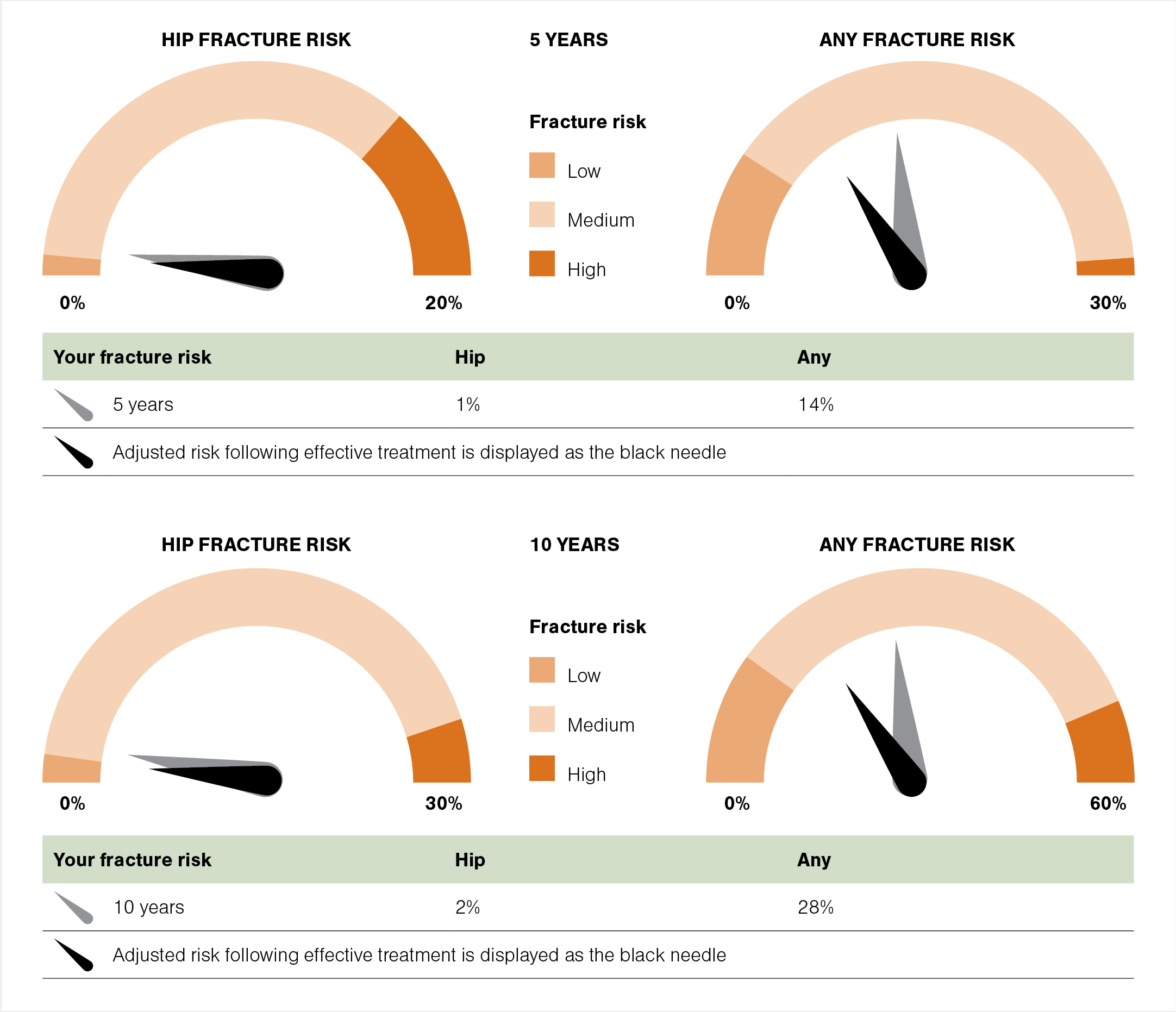
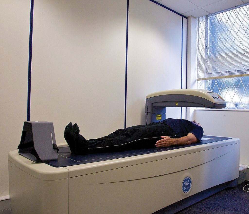
Regardless of whether there has been a fragility fracture or not, those at sufficiently high risk of future fractures should be considered for pharmacotherapy (a 10-year hip fracture risk above 3 per cent, using the FRAX or Garvan calculators, is often used as the threshold). This would include the majority of those having had any fragility fracture, especially those over age 75. This is the reason why a DXA scan is generally not necessary prior to initiating anti-osteoporosis treatment in this cohort. In particular, those with vertebral or hip fractures (associated with the highest risk of further fractures) should have lower threshold for treatment initiation after the sentinel fracture.

In the absence of fracture, treatment is also generally indicated in those whose DXA T-score is less than or equal to -2.5, or less than or equal to -1.5 with significant risk factors such that their fracture risk is high.

Oral and intravenous bisphosphonates

Bisphosphonates remain the first line and mainstay of anti-osteoporosis treatment, both locally and internationally. The ANZFFR Annual Report 2024 shows 95 per cent of anti-osteoporosis treatment used is either oral or intravenous bisphosphonate. Oral agents available in New Zealand are alendronate (Fosamax and Fosamax Plus) and risedronate, and the intravenous agent for use in osteoporosis is zoledronate. In the past, alendronate and zoledronate were funded under Special Authority restrictions, but both are now fully funded without restriction.

Studies suggest poor adherence to oral bisphosphonates. Therefore, it is recommended that adherence is checked periodically and that a blood test for procollagen-1 N-terminal peptide (P1NP) is performed about six months after starting treatment. P1NP is a bone formation (and hence turnover) marker that is suppressed (below 35µg/L) when oral bisphosphonate is taken regularly and absorbed adequately. Switching to intravenous zoledronate should be considered if P1NP is above 35µg/L despite seemingly good adherence or where consistent adherence is problematic.

Zoledronate infusion has the advantage that it only needs to be administered every 18 to 24 months, resulting in better patient acceptance and adherence. It can be used in patients who experienced side effects from, or have contraindications to, oral bisphosphonates.

A very common adverse effect of zoledronate infusion is post-dose flu-like symptoms that occur in up to one-third of patients after the first dose. This acute-phase response typically occurs in the first few days of drug infusion and resolves within three days or so. Paracetamol with or without an NSAID can be used to alleviate symptoms (the latter only if safe to use for the individual patient and deemed necessary). Incidence of this post-infusion adverse effect markedly decreases with subsequent infusions.

A recent New Zealand study showed that a three-day course of oral dexamethasone (4mg daily, started on the day of infusion) significantly reduced the acute-phase response (J Bone Miner Res 2023;38[5]:631–38). This approach could be considered either routinely or in those with significant prior post-infusion acute-phase response.

With the recent funding change for zoledronate, the fully funded product is no longer the well-known Aclasta but the generic version Zoledronic Acid Viatris. Therefore, we should avoid prescribing it as Aclasta, but instead prescribe zoledronate or zoledronic acid.

There remains an access inequality issue due to the zoledronate infusion fee that is charged by private and primary care providers. Due to the sheer volume of patients treated with zoledronate, secondary care in most regions of New Zealand is unable to offer infusions for all. There is an ongoing push by Osteoporosis New Zealand and Fracture Liaison Network New Zealand towards equitable and improved access for zoledronate infusions nationally.

Contraindications and adverse effects

Contraindications to bisphosphonates include renal failure – denoted in Medsafe data sheets as creatinine clearance (CrCl) <35ml/min for zoledronate and oral bisphosphonates. It is common practice to reduce the administration dose of zoledronate (eg, from 5mg to 2.5mg) and/or slow the infusion rate (over 30 to 60 minutes) when treating patients with a lower CrCl of 35–50ml/min.

Use of zoledronate in those with lower renal functions (eg, CrCl 25–35ml/min) and use of estimated glomerular filtration rate instead of CrCl remain controversial. Lower renal function threshold – possibly CrCl down to 15ml/min – for oral bisphosphonates is believed to be safe. Evidence for safety of this off-label use for oral bisphosphonates in stage 4 chronic kidney disease is scant, and hopefully will not be necessary once the access of denosumab (discussed in the next section) improves.

Oral bisphosphonates should also be avoided in those with significant impairment or delay in oesophageal emptying, such as oesophageal stricture or achalasia.

A well-publicised side effect of bisphosphonates is osteonecrosis of the jaw (ONJ). It manifests as an area of exposed bone in the mouth that does not heal within eight weeks. In reality, it is extremely rare in the setting of osteoporosis management. It is, nevertheless, recommended that significant dental issues requiring major dental work (eg, dental implants, multiple teeth extractions or jaw surgery) are resolved before initiation of bisphosphonate therapy.

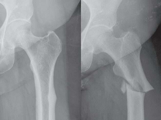
Another bisphosphonate-related long-term adverse effect is that of atypical femoral fracture (AFF). These initially begin as stress fractures in the lateral cortex of the femoral shaft and can spontaneously progress to full-thickness transverse fractures of the femur. Before fracture, the lateral cortex of the femur may appear thickened on x-ray, with a “beaked” appearance (Figure 3).

The incidence of AFF, albeit very low in absolute risk terms (several cases per 100,000 person-years), appears to increase steeply with increasing duration of bisphosphonate use, largely with oral forms, beyond five to seven years of treatment, and risk drops off dramatically within one to two years of treatment intermission or cessation. Thus, it is important to periodically review the need for continued bisphosphonate therapy and provide “drug holidays” (discussed below) for patients requiring therapy for more than five years.

Figure 3. X-rays taken several months before (left; note the “beaking” on the lateral cortical bone of the femur) and immediately after (right) an atypical femoral fracture

[Image: David Kim]

It is important to…provide “drug holidays” for patients requiring therapy for more than five years

Long-term follow-up

As alluded to above, it is recommended that the serum P1NP level be checked about six months after initiation of oral bisphosphonate therapy. P1NP can be checked at any stage of treatment if adherence and/or efficacy is being questioned, although there are certain situations where P1NP will not be reliable (eg, within a few months of a fracture). Total duration of bisphosphonate therapy and drug holidays are frequently debated and remain contentious issues.

Most patients established on bisphosphonate therapy should have a repeat DXA scan after four to five years of treatment. If the T-score has improved to -2.5 or higher without a recurrent fracture, treatment should be ceased for three to five years before reassessing fracture risk.

On the other hand, treatment should be continued for a further four to five years if the T-score remains less than -2.5. During this period, a one to two-year drug holiday should be considered if staying on oral bisphosphonates, to minimise the risk of AFF. For those on zoledronate infusion, the dosing interval could be increased to 24–30 months for the same reason, although AFF risk seems much lower in patients treated with zoledronate.

There is still limited evidence to guide bisphosphonate therapy (or any other anti-osteoporosis medication) beyond 10 years. Individuals with persistently high fracture risk should be considered for continued medical therapy with drug holidays every several years, and guidance from secondary care could be sought. Similarly, patients with recurrent fractures despite adequate therapy, or those with intolerances and/or contraindications to both oral and intravenous bisphosphonates, should be referred to secondary care.

Increasing use of non-bisphosphonate anti-osteoporosis medications

Some of these agents are simply superior in terms of anti-fracture efficacy

There has been a recent secular trend of increasing use of non-bisphosphonate medications for osteoporosis, although they still only make up 5 per cent of treatments used, according to the ANZFFR Annual Report 2024. However, their use is expected to continue to increase with likely easing of Pharmac funding restrictions in coming years and with increasing familiarity and experience of prescribers using non-bisphosphonate agents.

These agents can overcome issues that bisphosphonates may have, and some agents are simply superior in terms of anti-fracture efficacy.

Oestrogen-based therapies

Hormone replacement therapy, these days referred to as menopausal hormone therapy, incorporates oral or transdermal oestrogen. Once deemed unsafe in all postmenopausal women, MHT has made a noticeable comeback in recent years. There have been reappraisals of the evidence to suggest the risk–benefit profile has moved significantly in favour of its use, including for preventing and treating osteoporosis or high fracture risk states in younger menopausal women.

In the original Women’s Health Initiative study, women on HRT were found to have lower fracture risk, and other studies also consistently show favourable bone effects of MHT. Safety in women within the first 10 years of menopause has been well demonstrated in a number of studies, and MHT could be used as a second-line agent in women under age 60 (sometimes even a little above this age threshold) who do not have additional risk factors for cardiovascular disease, breast cancer or venous thromboembolism.

Oral forms of oestrogen, such as conjugated oestrogen and oestradiol valerate, can be used. The transdermal preparation fully funded in New Zealand is the oestradiol patch (Estradot or Estradiol Transdermal System). Oral oestrogens are perhaps more convenient, though the transdermal route theoretically carries a lower side-effect risk, bypassing first-pass metabolism.

Regardless of whether oral or transdermal oestrogen is used, in those with an intact uterus, a progestogen should be used for endometrial protection – cyclical or continuous medroxyprogesterone (Provera) or micronised progesterone (Utrogestan).

Risk versus benefit of MHT should be considered before treatment initiation.

Contraindications to oestrogen therapy include history of ischaemic heart disease, breast cancer or sex hormone-responsive tumours, liver tumours, venous thromboembolism and severe liver disease. MHT use in those with significant cardiovascular risk factors should also be avoided.

Dosing information for oestrogen and corresponding progestogen is well summarised in the Australasian Menopause Society’s AMS Guide to MHT/HRT Doses New Zealand Only (tinyurl.com/MHT-doses).

Teriparatide

Teriparatide (Forteo) is a synthetic parathyroid hormone (PTH 1–34) analogue that has been available in New Zealand for many years. Given as a daily subcutaneous injection (20μg), it stimulates osteoblast activity and acts as an anabolic agent for bone.

Current funding criteria (Panel 3) is quite restrictive, in that patients only qualify if they have had two or more fragility fractures, at least one of which after having had adequate bone protection therapy (bisphosphonate or raloxifene for more than one year), and with very low bone mineral density (T-score less than -3.0). Any relevant practitioner can apply for the Special Authority. There is a compelling argument for its use as a first-line agent in very severe osteoporosis. Pharmac is currently reconsidering teriparatide funding.

Teriparatide comes in a pre-filled injection device that patients need to learn to self-inject. Often, drug initiation is done in secondary care, although many primary care practices are now initiating therapy independently

The approved duration of use for teriparatide is 18 months in New Zealand (up to 24 months in some other countries), and it is not to be used in conjunction with bisphosphonates. Bisphosphonate therapy, usually in the form of intravenous zoledronate, is recommended at the end of the 18-month treatment course.

PANEL 3. Pharmac Special Authority criteria for teriparatide

Patient has severe, established osteoporosis.

AND

The patient has a documented T-score less than or equal to -3.0 (must be made using DXA).

AND

The patient has had two or more fractures due to minimal trauma.

AND

The patient has experienced at least one symptomatic new fracture after at least 12 months’ continuous therapy with a funded antiresorptive agent at adequate doses (alendronate 70mg or 70mg with cholecalciferol 5600IU once weekly; raloxifene 60mg once daily; zoledronate 5mg per year).

Denosumab

Denosumab (Prolia) is available as a six-monthly subcutaneous injection. It is a potent antiresorptive agent that works in a similar fashion to the bisphosphonates but via a different cellular pathway and with superior anti-fracture efficacy. Denosumab is not cleared renally; therefore, it can be used safely in those with moderate to severe (stages 3 and 4) chronic kidney disease.

Denosumab has been available in New Zealand for several years and remains under highly restrictive funding criteria that include the presence of severe osteoporosis with recurrent fracture despite being on adequate antiresorptive therapy for over one year and CrCl <35ml/min (Panel 4). Efforts to broaden funding indications for denosumab over the years have been unsuccessful, although it is hoped that criteria will relax in coming years. Drug cost (generally under $1000 per year) is not overly prohibitive for many patients, and an increasing number of patients with severe osteoporosis are choosing to self-fund denosumab.

In patients receiving denosumab, ONJ and AFF have been described but are very rare, and there are long-term (10-year) safety and efficacy data. It has been shown to be superior to bisphosphonates in terms of bone density gains and anti-fracture efficacy, and its long-term efficacy is particularly impressive.

Denosumab could serve as a second-line agent for patients who are intolerant of, or refracture while on, bisphosphonate therapy. Easy and infrequent administration makes it an attractive treatment option, especially in patients in whom administration of bisphosphonates poses difficulty.

Arguably the most important drawback of denosumab is the well-described phenomenon of rapid offset of drug effect if discontinued or if dosing is significantly delayed. Especially in those who have been on denosumab therapy for more than three years, a dramatic rise in bone turnover markers and rapid fall in bone density are observed after treatment cessation, with significant increase in vertebral compression fractures.

The importance of continuation of therapy with regular six-monthly dosing cannot be overemphasised. For this reason, denosumab treatment is often considered in relatively older patients with severe osteoporosis who are committed to taking it for the rest of their life. If denosumab is to be ceased, the case must be discussed with a secondary care colleague with relevant experience and expertise.

PANEL 4. Pharmac Special Authority criteria for denosumab

Patient has severe, established osteoporosis.

AND

  • The patient is female and postmenopausal. OR
  • The patient is male or non-binary.

AND

  • History of one significant osteoporotic fracture demonstrated radiologically and a documented T-score less than or equal to -2.5 (must be made using DXA). OR
  • History of one significant osteoporotic fracture demonstrated radiologically, and either the patient is elderly or densitometry scanning cannot be performed because of major logistical, technical or pathophysiological reasons. OR
  • History of two significant osteoporotic fractures demonstrated radiologically. OR
  • Documented T-score less than or equal to -3.0 (must be made using DXA). OR
  • A 10-year risk of hip fracture ≥3 per cent, calculated using a published risk assessment algorithm (eg, FRAX or Garvan) which incorporates DXA bone mineral density measurements. OR
  • Patient has had a Special Authority approval for alendronate (underlying cause – osteoporosis) prior to 1 February 2019 or has had a Special Authority approval for raloxifene.

AND

Zoledronic acid is contraindicated because the patient’s CrCl is <35ml/min.

AND

The patient has experienced at least one symptomatic new fracture after at least 12 months’ continuous therapy with a funded antiresorptive agent at adequate doses (risedronate 35mg once weekly; alendronate 70mg or 70mg with cholecalciferol 5600IU once weekly; raloxifene 60mg once daily).

AND

The patient must not receive concomitant treatment with any other funded antiresorptive agent for this condition or teriparatide.

Raloxifene

Raloxifene (Evista) is a daily oral therapy for osteoporosis that is available under relatively loose funding criteria, which includes DXA, clinical fracture or fracture risk criteria. Raloxifene is rarely used for osteoporosis, largely due to a lack of anti-fracture efficacy for non-vertebral fractures as well as a side-effect profile that includes flushing.

Romosozumab

This potent, highly effective anti-osteoporosis agent has both bone anabolic and antiresorptive properties. It is delivered as a monthly subcutaneous injection for one year only. Romosozumab has recently become available in Australia under strict funding restrictions but is not yet available here.

Referral to secondary care

All regions in New Zealand have secondary care services that can provide advice or review patients with difficult to manage osteoporosis. Subspecialties playing this role vary by region and are typically endocrinology, older people’s health/orthogeriatrics, and rarely rheumatology, orthopaedic surgery and primary care (GP with special interest). Many of these clinicians will be involved with their local FLS and provide clinical leadership and/or be involved in regional DXA services.

Referral threshold varies regionally, but the following cases should be considered for referral:

  • recurrent fragility fracture despite appropriate anti-osteoporosis medication
  • intolerance to, or strong aversion to, standard anti-osteoporosis medication (ie, oral or intravenous bisphosphonate with or without MHT)
  • “idiopathic” osteoporosis (Z-score worse than -2.0) in those under the age of 50
  • patients who have received 10 years or more of bisphosphonate therapy and remain at high risk of fracture
  • those on denosumab who may need to discontinue therapy fracture
  • any other situation where the use of teriparatide or denosumab may be indicated.
Patient information

Bone Health New Zealandbones.org.nz/fact-sheets

Osteoporosis New Zealandosteoporosis.org.nz

UpToDate. Patient education: Osteoporosis prevention and treatment (Beyond the Basics)tinyurl.com/patients-osteo

Live Stronger for Longerlivestronger.org.nz

International Osteoporosis Foundationosteoporosis.foundation/patients

Further reading and resources

Osteoporosis New Zealand. Guidance on the Diagnosis and Management of Osteoporosis in New Zealand. 2017tinyurl.com/guidance-osteo

Osteoporosis New Zealand. Clinical Standards for Fracture Liaison Services in New Zealand, 2nd edition. 2021 tinyurl.com/stds-fls

Australasian Menopause Society. AMS Guide to MHT/HRT Doses New Zealand Only. 2023tinyurl.com/mht-doses

Regional HealthPathwayshealthpathwayscommunity.org

CASE STUDY 1. Drastic weight loss a significant risk factor for bone loss

Presentation and history

A 63-year-old woman used to be “overweight” with a BMI in the obese range (>40kg/m2), but in the context of regular high-intensity resistance training at the gym. She experienced a T10 vertebral compression fracture after “a very heavy fall” five years ago, with a DXA scan at the time showing only mild osteopaenia, and no anti-osteoporosis treatment was implemented.

She has lost about 50kg of weight since then (current BMI 26kg/m2). This has largely been intentional through dietary changes as well as from loss of muscle mass after stopping resistance training – the latter due to back pain following the fracture as well as significant degenerative changes needing orthopaedic interventions.

A repeat DXA scan was recently performed, principally because of the drastic weight loss and known osteopaenia. This shows significant reduction in bone mineral density compared with the previous scan, with osteoporosis in the lumbar spine (T-score -2.8) and advanced osteopaenia in average total hips (T-score -2.3). Calculated fracture risk confirms increased hip fracture risk (3.6 per cent with FRAX New Zealand and 7 per cent with Garvan).

After discussing these results with the patient, a zoledronate infusion is delivered (5mg over 15 minutes), with a plan for two further infusions every 18 months. She has plans to get back into resistance training, as back pain allows.

Learning points and follow-up

This case illustrates the importance of looking out for significant bone loss in someone with concurrent significant risk factors (drastic weight loss and pre-existing osteopaenia in this case). Implementation of treatment (zoledronate infusion in this case) is expected to improve bone mineral density by several percentage points over the first three to five years, with overall fracture risk reduction close to 50 per cent.

A DXA scan can reasonably be performed a year or so after the third infusion of zoledronate to help make a decision about either continued zoledronate treatment or three to five years of a drug holiday.

Dual-energy x-ray absorptiometry is the recommended diagnostic test for osteoporosis [Image: Nick Smith photography, CC BY-SA 3.0]
CASE STUDY 2. Do not cease denosumab abruptly

Presentation and history

A 64-year-old woman was diagnosed with rheumatoid arthritis eight years ago when she was living in Australia. She needed to take supraphysiological doses of prednisone in the early stages of managing her arthritis. Despite implementation of various disease-modifying agents, including biologics, she still takes prednisone 5mg daily.

She had a pubic rami fracture five years ago in Australia, and a DXA scan reportedly showed significant osteoporosis. Denosumab (Prolia) was initiated, and six-monthly injections were continued up until moving to New Zealand nearly a year ago. Her last DXA scan was performed in Australia about 18 months ago – result not available but explained by the Australian physician as being “okay”.

Denosumab was not continued after coming to New Zealand; she was told by her primary care provider that denosumab was “not available” in New Zealand, and no anti-osteoporosis treatment has been administered since.

About three months after she was due for her denosumab injection, she presented with unprovoked back pain, with subsequent imaging confirming multi-level thoracolumbar vertebral compression fractures (see CT scan). Back pain has been quite severe, such that she has had to stop working, needs regular analgesic agents in combination, including opioids and pregabalin, and needs a walking frame to mobilise.

Diagnosis, management and follow-up

This woman with significant pre-existing risk factors for fragility fractures experienced multi-level vertebral fractures due to abrupt cessation of denosumab therapy.

During the first bone clinic review, she is given the option of either switching over to a bisphosphonate or reinitiating denosumab – the latter is not funded in this case. She opts to self-fund denosumab.

DXA is performed a few months later and shows osteopaenic-range results, with a T-score of -1.5 in the total hips and -1.9 in the neck of the femur (unable to scan spine due to compression fractures). She has had no further fractures. Six-monthly denosumab will be continued indefinitely.

Learning points

Anyone having been treated with denosumab, especially if the treatment duration was over two years, must not have their treatment ceased abruptly. If treatment cessation is being considered, immediate discussion with, or referral to, secondary care is imperative.

CT scan showing multi-level vertebral compression fractures [Image: David Kim]

Details have been changed to protect patient confidentiality

David Kim is an endocrinologist and general physician at Te Whatu Ora Waitematā and Apollo Health & Wellness, Auckland

Assessment

This 10-question multiple-choice assessment is designed to demonstrate that the provided educational reading has been effective in allowing you to meet the learning objectives of this course. Write down your answers to these questions.

1. Which two statements regarding osteoporosis are correct?
a.
Approximately three-quarters of those affected are women
b. As we age, osteoblast activity outpaces that of osteoclasts
c. Osteoporosis incidence is equal across all ethnicities in New Zealand
d. Well over 20,000 New Zealanders experience osteoporosis-related fractures each year

2. What roles do primary care play in managing osteopaenia, osteoporosis and fragility fractures?
a.
Screening and evaluation of all patients potentially at risk for low bone density
b. Implementation of lifestyle modifications and falls prevention strategies
c. Assessment of fractures seen in primary care, including determining whether bone fragility is a factor
d. Follow-up and management of patients treated for fragility fractures in secondary care
e. Initiation, continuation and follow-up of anti-osteoporosis treatment
f. All of the above

3. Which two of the following statements concerning dual-energy x-ray absorptiometry are correct?
a.
A DXA scan is generally not necessary prior to initiating anti-osteoporosis treatment for those over age 75 with a fragility fracture
b. DXA is the best validated tool for diagnosing osteoporosis and assessing fragility fracture risk
c. DXA requires a moderate dose of radiation
d. There is low inter-regional variability in access to funded DXA scans in New Zealand

4. When interpreting the results of a DXA scan, which two of the following statements are correct?
a.
A T-score between zero and -2.49 represents osteopaenia
b. A T-score of -2.5 or less represents osteoporosis
c. People with a Z-score below -2.0 should be assessed for secondary causes of bone loss regardless of age
d. T-score is the gender and age-matched standard deviation score of bone density

5. At what point should patients generally be offered pharmacotherapy to reduce the risk of fragility fracture (three correct answers)?
a.
DXA T-score -2.5 or less
b. DXA T-score -1.5 or less
c. DXA T-score -1.5 or less with significant risk factors
d. DXA Z-score -2.0 or less in those under age 50
e. FRAX or Garvan calculator 10-year hip fracture risk >1.5%
f. FRAX or Garvan calculator 10-year hip fracture risk >3%

6. Which two statements regarding intravenous zoledronate are correct?
a.
A three-day course of oral dexamethasone 4mg daily reduces the acute-phase response associated with zoledronate infusion
b. Risk of atypical femoral fracture is higher in patients treated with zoledronate than oral bisphosphonates
c. Switching from oral bisphosphonate therapy to intravenous zoledronate should be considered if the procollagen-1 N-terminal peptide level is above 35μg/L
d. Zoledronate infusion has the advantage that it only needs to be administered every six to nine months
e. Zoledronate should be prescribed as Aclasta

7. Contraindications for bisphosphonates include which two of the following?
a.
Creatinine clearance <35ml/min
b. Estimated glomerular filtration rate <45ml/min/1.73m2
c. Inflammatory bowel disease
d. Significant impairment in oesophageal emptying

8. Bisphosphonates do have adverse effects – which two of the following statements concerning them are correct?
a.
Osteonecrosis of the jaw is very rare in the setting of osteoporosis treatment
b. Risk of atypical femoral fracture is constant throughout treatment
c. Risk of atypical femoral fracture remains raised indefinitely following treatment cessation
d. Significant dental issues should ideally be resolved before bisphosphonate treatment

9. Patients established on oral bisphosphonate therapy need which two of the following?
a.
P1NP level checked about six months after treatment initiation
b. Repeat DXA scan after one to two years
c. Treatment cessation if no recurrent fracture after four to five years, regardless of the T-score
d. Consideration of “drug holidays” beyond five years of therapy

10. For fragility fracture patients adhering well to oral bisphosphonate therapy for more than 12 months (P1NP level <35μg/L) and with a T-score -3.0 or less, who then have a second fragility fracture while on treatment, what two other treatment options are available and advisable to consider?
a.
Denosumab (funded only for CrCl <35ml/min)
b. Raloxifene
c. Romosozumab
d. Teriparatide
e. Zoledronate

Write down your answers to these questions. Then, to check your answers and record your score, click here.

Thinking about learning?

You can use the Capture button below to record your time spent reading and your answers to the following learning reflection questions, which align with Te Whanake reflection requirements (answer three or more):

  • What were the key learnings from this activity?
  • How does what you learnt benefit you, or why do you appreciate the learning?
  • If you apply your learning, what are the benefits or implications for others?
  • Think of a situation where you could apply this learning. What would you do differently now?
  • If an opportunity to apply this learning comes up in the future, what measures can be taken to ensure the learning is applied?
  • Can you think of any different ways you could apply this learning?
  • Are there any skills you need to develop to apply this learning effectively?
FREE and EASY

We're publishing this article as a FREE READ so it is FREE to read and EASY to share more widely. Please support us and our primary care education resources – subscribe here

PreviousNext