The ubiquitous mole – almost everyone has them, so it pays to know their subtleties

FREE READ
+Summer Hiatus
In print
FREE READ

The ubiquitous mole – almost everyone has them, so it pays to know their subtleties

ELearning now available for doctors

Amanda Oakley

5 minutes to Read
Dermatoscopic examination 1
Dermatoscopic examination

We are on our summer break and the editorial office is closed until 17 January. In the meantime, please enjoy our Summer Hiatus series, an eclectic mix from our news and clinical archives and articles from The Conversation throughout the year. This article was first published in the 12 May edition

Melanocytic naevi can be classified into three main groups according to where naevus cells are found in the skin. In this article, dermatologist Amanda Oakley describes their appearance and dermatoscopic features

Key Points
  • Naevi mainly arise in childhood or adolescence and may slowly evolve.
  • Most naevi appear symmetrical in colour and structure.
  • Dermatoscopy shows regular reticular, clod or structureless patterns, or symmetrical combinations of these.

This article has been endorsed by the RNZCGP and has been approved for up to 0.25 CME credits for the General Practice Educational Programme and continuing professional development purposes (1 credit per learning hour). To claim your credits, log in to your RNZCGP dashboard to record this activity in the CME component of your CPD programme.

Nurses may also find that reading this article and reflecting on their learning can count as a professional development activity with the Nursing Council of New Zealand (up to 0.25 PD hours).

Melanocytic naevi, or moles, are easily recognisable with some basic knowledge, clinical examination and the use of a dermatoscope. Some naevi are trickier and we expect these to be referred or excised when there is high suspicion of malignancy.

Melanocytic naevi are due to the benign proliferation of naevus cells (specialised melanocytes). They are thought to arise after one or more cellular mutations.

Most naevi appear before puberty, but they may arise during adolescence or early adult life. Almost everyone has one or more, and some people have hundreds of them. Moles occur in all skin types, light and dark, but are more numerous and variable in appearance in light skin, following sun exposure and in people who are genetically predisposed to them. They tend to be small (<6mm), symmetrical in colour and structure, and regular in outline. Any change due to age, sun exposure or immune-related factors usually occurs slowly. Some naevi are large, with variable colour and structures, but remain stable or change very slowly.

Naevi often resemble others on the same individual and may be distributed along embryonic lines. In Caucasian people, they are most common on the head, neck and trunk; in Asian people, they are most common on palms and soles.

Classification of melanocytic naevi

Junctional naevi – these naevi get their name from the site of nests of naevus cells or lentiginous proliferation within the epidermis. They are acquired as a response to sun exposure and can arise at any age on the upper trunk and proximal limbs. They are flat, tan or brown marks and may have a darker centre or variation in colour distribution (Figures 1a,2a and 3a). In later years, junctional naevi may involute by fading and shrinking.

Dermal naevi – the melanocytic proliferation in a dermal or intradermal naevus is within the dermis. They arise before the age of 10 to 15 on any site of the body. They can look like small birthmarks (Figure 4a), and some bear terminal hair (Figure 5a). Over time, dermal naevi may become larger and thicker, gaining or losing colour. A papule on the trunk in childhood (Figure 6a) may enlarge in adulthood to become pedunculated with a papillomatous surface (Figure 7a). On the face and neck, dermal naevi tend to be firm with a smooth surface. They are pink, red, light brown, dark brown or black, depending on skin colour (Figures 8a, 9a and 10a).

Compound naevi – these naevi have junctional and dermal components (Figures 11a, 12a and 13a).

Dermatoscopic examination

Examination of a naevus with a dermatoscope allows an evaluation of the global pattern and focal features.
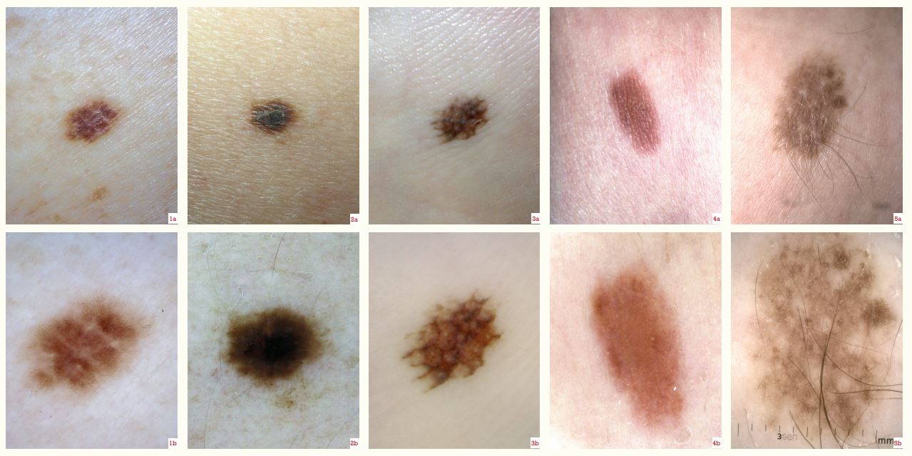
Junctional naevi are recognised by a reticular pattern of lines in most areas (Figures 1b and 2b) and by parallel lines on the palms and soles (Figure 3b), or they appear structureless. The lines fade peripherally.

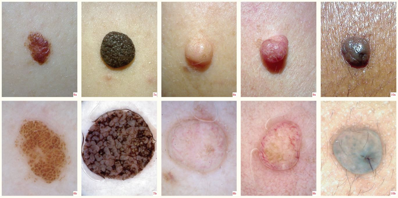
Dermal naevi – are structureless (Figures 4b, 8b, 9b and 10b) or are made up of clods, which are small and separated (Figure 6b), or large (Figure 7b). Keratin plugs may be seen as orange/brown, irregularly distributed, superficial clods (Figure 7b). Facial, non-pigmented dermal naevi have faint to prominent, curved or serpiginous, non-branched vessels (Figures 8b and 9b).

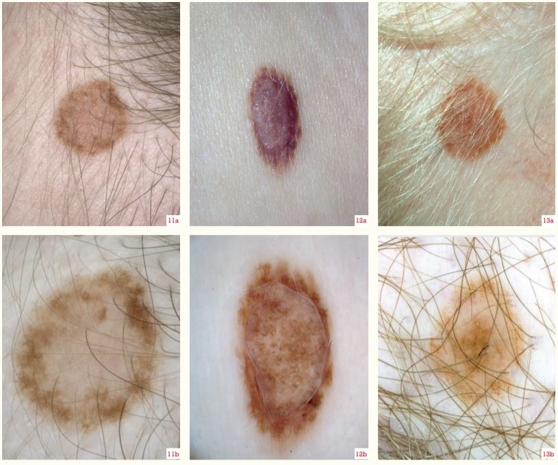
Compound naevi – have both features, symmetrically distributed as two patterns (Figures 11b and 12b) or three concentric patterns (Figure 13b). The dermal component may not be clearly seen (Figure 5b; in this example, perifollicular hypopigmentation and thick hair growth indicate the naevus is of congenital pattern and will have a dermal component).

Naevi have particular dermatoscopic characteristics according to site, especially noticeable on the scalp (Figures 11b and 13b; an annular pattern is common), face (Figures 8b, 9b and 10b), and palm/sole (Figure 3b; a parallel furrow pattern, best appreciated on the lesion edge).

Further investigation

Naevi are harmless and do not need to be removed; if they are of clinical concern, diagnosis requires referral for another opinion or complete excision with a 2mm clinical margin of uninvolved skin. Pathological examination of a naevus subjected to biopsy may show:

  • small, round melanocytes with uniformly stained nuclei and scant cytoplasm
  • nested proliferation at the tips of the rete ridges (these are absent in lentigines); lentiginous naevi are arranged as single cells along the base of the epidermis
  • dermal melanocytes decreasing in size towards the base of the lesion in strands or chords
  • a congenital growth pattern that involves adnexae or a reticular dermis
  • site-related features
  • absent or rare mitoses, necrosis or cytological atypia
  • specific cytogenetic features. In an irritated naevus, signs of recent trauma, including ulceration and inflammation, may be observed.
The images show the following conditions:

Figure 1a. An acquired junctional naevus
Figure 1b. Dermatoscopy of 1a shows a reticular pattern
Figure 2a. Junctional naevus with a dark, scaly centre
Figure 2b. Dermatoscopy of 2a shows a dark, structureless centre and reticular periphery
Figure 3a. Acral naevus
Figure 3b. Dermatoscopy of 3a shows a parallel furrow pattern with some lattice pattern centrally
Figure 4a. A solid-brown dermal naevus
Figure 4b. Dermatoscopy of 4a shows it to be structureless and brown
Figure 5a. Congenital-like naevus with terminal hair growth
Figure 5b. Dermatoscopy of 5a shows a reticular pattern and perifollicular hypopigmentation

Figures 1-5 (click on red arrow to enlarge image)

Figure 6a. Common naevus in childhood
Figure 6b. Dermatoscopy of 6a shows small brown clods
Figure 7a. Papillomatous dermal naevus
Figure 7b. Dermatoscopy of 7a shows a large brown clod pattern with keratin in the surface crevices
Figure 8a. Skin-coloured, dome-shaped, facial dermal naevus
Figure 8b. Dermatoscopy of 8a shows it to be mainly structureless with flecks of pigment and faint, curved, linear blood vessels
Figure 9a. Pink, facial dermal naevus
Figure 9b. Dermatoscopy of 9a shows localised, structureless pigmentation and prominent serpiginous vessels
Figure 10a. Dermal facial naevus in dark skin
Figure 10b. Dermatoscopy of 10a shows hazy and patchy pigmentation and a central terminal hair

Figures 6-10 (click on red arrow to enlarge image)

Figure 11a. Annular scalp naevus
Figure 11b. Dermatoscopy of 11a shows the pigmented periphery is reticular with a structureless, non-pigmented centre
Figure 12a. Compound naevus – the dermal naevus has acquired peripheral pigment
Figure 12b. Dermatoscopy of 12a shows a peripheral network and central, patchy pigmentation
Figure 13a. Targetoid or cockade scalp naevus
Figure 13b. Dermatoscopy of 13a shows it to have three concentric zones

Figures 11-13 (click on red arrow to enlarge image)

Amanda Oakley, CNZM, is a specialist dermatologist at Waikato Hospital, an adjunct associate professor at Waikato Clinical Campus, and founder of DermNetNZ.org

Thinking about learning

You can use the Capture button below to record your time spent reading and your answers to the following learning reflection questions:

  • Why did you choose this activity (how does it relate to your PDP learning goals)?
  • What did you learn?
  • How will you implement the new learning into your daily practice?
  • Does this learning lead to any further activities that you could undertake (audit activities, peer discussions, etc)?
FREE and EASY

We're publishing this article as a FREE READ so it is FREE to read and EASY to share more widely. Please support us and our journalism – subscribe here

PreviousNext