MARS Bioimaging Ltd (MBI) will soon begin a large-scale international trial of its revolutionary new compact 3D-colour hand and wrist scanner, following a successful feasibility study involving New Zealand orthopaedic patients.
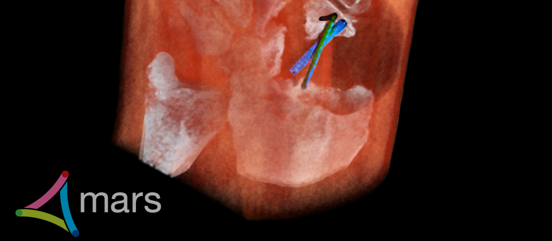
The feasibility study produced the first ever patient images from the world-first compact scanner developed especially to better diagnose hand and wrist problems.
MBI invented the world’s first 3D colour scanner using technology adapted from the European Organization for Nuclear Research (CERN) in Switzerland and its Large Hadron Collider. In 2018, after more than a decade of research in partnership with the universities of Canterbury and Otago, the MBI team scanned the first living body.
They have since developed a specialist compact scanner for diagnosing hand and wrist injuries and will begin clinical trials from early 2021, including with patients from New Zealand’s largest radiology service provider, Pacific Radiology Group.
President Medical Professor Anthony Butler says a feasibility study of the hand and wrist scanner by radiologists and surgeons in 2019 found it provided significant improvements in the diagnosis of hand and wrist fractures compared with existing technology. Read a summary of the feasibility study.
“Results from our initial patient trials showed high-resolution spectral imaging could provide significant improvements in the diagnosis of hand and wrist injuries. We chose to target the important clinical problem of wrist injuries because they are common and diagnosis can be challenging, with frequent misdiagnosis and complications such as bones not healing properly”, says Professor Butler.
“Unparalleled clarity means diagnosing complications, such as minute fractures that aren’t healing correctly, is dramatically improved. The MBI scanner’s ability to measure and display bone composition makes it far easier to monitor post-surgical healing. It can also focus on the optimal energy spectrum to reduce image distortion caused by metal implants and better assess bone healing and fusion.”
Following the results of the feasibility study, Professor Butler says MBI will soon begin extensive testing of its wrist scanner at Pacific Radiology Group’s 24-hour surgery clinic in Christchurch, New Zealand. Pacific Radiology Group is a key collaborative partner on this ground-breaking research project. MBI will also expand trials internationally by installing a wrist scanner in a clinic at Lausanne University Hospital in Switzerland within the next few months.
“This technology is really out there”, says Dr Ross Keenan, Pacific Radiology Head of Research and Development.
“A detector chip from CERN is very novel and it’s potentially the way of the future. We are aiming for more and more information with virtually no dose of radiation at all. The exciting thing is this technology has been produced locally and it could completely transform the way we do point of care imaging. There is nothing else like it.”
Aurélie Pezous, Knowledge Transfer Officer at CERN says: “For more than twenty years, CERN has been putting its know-how in the field of hybrid pixel detectors at the service of medical innovation. Spectroscopy imaging is at the heart of Medipix3 chips, allowing for X-ray colour imaging. The first clinical trials represent a great step for this technology as well as a source of pride for CERN and the Medipix3 Collaboration.”
MBI’s wrist scanner was developed as a compact system for small clinics or surgeries to reduce delays and costs for healthcare providers and improve patient outcomes. The technology provides radiologists with all the details of a CT, but in high-definition colour and with information about tissue health and composition, normally only available with MRI and PET.
Pending regulatory approvals, MBI’s spectral wrist scanners could be available for clinical use within the next year, Professor Butler says. The wrist application is the first in a wide range of scanners planned for many areas of medicine.
Professor Butler says: “The use of Medipix technology in human trials represents a long-term investment by the New Zealand government in developing the country’s high-tech industry.”
MBI delivered the world’s first commercial Medipix-technology scanner for use in pre-clinical research. This scanner is now in use at dozens of prestigious universities across the globe.